Topic index: A
B C
D E
F G
H I
J K
L M
N O
P R
S T
U V
W X
Z
Procedures Treatments
Home
J
Juvenile
plantar dermatosis
Jessner lymphocytic infiltrate
Jessner’s lymphocytic infiltrate is a benign T-cell lymphocytic
infiltration of the skin that presents as non-scaly red patches
and lumps on the face, neck and upper back.
Cause
It is caused by a benigh proliferation of T-cell lymphocytes.
Why this happens is uncertain. Some cases have been associated
with borrelia infection, the cause of Lyme disease.
Signs
Red tumid papules, nodules or plaques. Lesions occasionally
have an arciform (arc-like) shape
The skin surface is smooth without scale or plugging.
Size varies from 2 mm to 2 cm
May be single or multiple.
Lesions enlarge gradually and in some cases clear in the centre
to give a annular (ring-like) or arciform (arc-like) shape.
Lesions may improve and worsen over months or years and seasonal
activity is variable with most patients experiencing more active
symptoms over the winter. Sometimes, they may resolve completely.
Diagnosis
A skin biopsy is necessary to confirm the diagnosis and exclude
other conditions such as cutaneous lymphoma, tumid lupus erythematosus,
lymphocytoma cutis, sarcoid, granuloma faciale and polymorphous
light eruption. It was believed that irt was in the same spectrum
as tumid lupus erythematosus monoclonal antibody Leu 8 staining
suggests that they are probably separate conditions.
Outlook
Harmless. May persist for months or years and resolve without
leaving scars or causing other problems. Recurrence may occur
at the same site or elsewhere.
What you can do
Photoprotection may help since lesions often arise on UV-exposed
sites, regardless of their history of photo-aggravation.
Treatment
Usually no treatment is necessary for Jessner lymphocytic infiltrate
as it usually resolves after periods of a few months to years,
Cosmetic camouflage may be used to hide lesions and improve appearance.
There is variable response to the following treatments:
Potent topical or intralesional steroids
Antimalarial medications, such as hydroxychloroquine, which has
an anti-inflammatory effect in the skin
UVA1 phototherapy or photochemotherapy (PUVA)
Photodynamic therapy
Cyclophosphamide
Thalidomide
Radiotherapy
JUVENILE PLANTAR DERMATOSIS (JPD)
This is a type of glazed fissured
dermatitis affecting the feet of children, especially boys between
7 - 12 years of age.
Causes
- JPD is believed to be caused
bt repeated friction inside footwear. The frictional force is
increased in sweaty feet which is why the the wearing of occlussive
footwear such has heavy winter shoes and synthetic shoes predisposes
to it.
- 'Atopic' children with atopic
dermatitis (eczema), asthma, or hay fever are predisposed to
JPD.
- Symptoms
- Glazed, cracked skin affecting
the forefeet and toe pads, sparing the toe webs and insteps.
- Affects both feet symmetrically.
- Itching is usually absent.
- Cracks may cause pain.
- The hands may sometimes be
affected, as well.
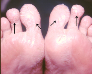 |
Juvenile plantar dermatosis.
Click on
image for larger view |
Complications
- Cracking and bleeding.
- Secondary bacterial infection.
What you can do
- You should consult a doctor.
- Wear well fitting shoes, preferably
leather
- Use cotton socks which allow
better ventilation.
- Moisturise the skin so it
is less likely to crack.
What the doctor may do
- Prescribe mild topical steroids and tar preparations.
- Exclude other causes such
as atopic dermatitis, contact dermatitis, psoriasis, keratolysis
exfoliativa, or a fungal infection
(see tinea
pedis).
- Perform patch tests to exclude
allergy to footwear.
TOP
Juvenile xanthogranuloma
(JXG)
JJuvenile xanthogranuloma (JXG)
is a rare type of non-Langerhan's cell histiocytosis (Class IIb)
that is benign. More common in children. This disease may have
been first reported by Rudolf Virchow in 1871 and again in 1905
by H.G. Adamson. In 1954, it was named juvenile xanthogranuloma
to reflect the appearance of the cells under a microscope.
Cause
- JXG is caused by the over-production
of a kind of histiocyte called a dendritic cell (not a macrophage)
but why this happens is not known.
Signs
- It presents as skin lesions
predominantly in infants and young children, more often males,
and is present at birth in 20% of cases. However, 10% of cases
are adults. It is more common in Caucasians than in those of
oriental origin.
- Smooth and pink papule, which
later develops a yellowish hue and may become scaly.
- Usually solitary but may be
multiple.
- Usually occur on the head,
neck and trubk but may occur anywhere.
- mainly affects infants and
small children with an average age of 2 years, although it can
also occur in adults of all ages. more males are affected than
females.
- However 20% also have café
au lait macules. Multiple café au lait birthmarks are
associated with neurofibromatosis type 1.
- JXG can affect the eye, most
commonly in young children with multiple skin lesions. Less commonly
JXG may involve locations such as the lung, liver, adrenal gland,
appendix, bones, bone marrow, pituitary gland, central nervous
system, kidney, heart, small and large intestines, and spleen.
Variants - Juvenile xanthogranuloma
is difficult to distinguish from several other conditions.
- Benign cephalic histiocytosis
- Usually occurs in infants less than one year of age. Presents
with multiple small reddish-brown bumps on the skin, mostly on
the head and neck. They are increasingly thought of as the same
condition.
- Generalised eruptive histiocytosis
- Usually affects adults. Presents with hundreds of reddish-brown,
blue-red or skin-coloured papules (less than 1cm) on the skin
of the trunk, face, upper arms and thighs. Recurrent crops of
lesions may appear and spontaneously resolve after several months.
May leave scars.
- Papular xanthogranuloma -
Occurs in children and adults. Papules occur in the skin and
in adults, in the lining of the mouth, Spontaneously resolves
in children but may persist in adults. There is no effective
treatment at this time
Diagnosis
The diagnosis of juvenile xanthogranuloma
and related conditions can be confirmed by a skin biopsy. The
lesions are composed of collections of histiocytes. In older lesions,
the histiocytes may appear foamy, filled with lipids (fats) or
hugely enlarged (giant cells).
Treatment
- Skin lesions are self-limited
and rarely require treatment in most patients.
- Those with large abdominal
masses, liver, bone marrow, or central nervous system involvement
may do well with treatment such as chemotherapy similar to that
used for Langerhans cell histiocytosis. Because this disease
is so rare, no large studies have been performed, and there is
no established, proven treatment for the more complicated cases.
- Unless they occur in the eyes,
juvenile xanthogranulomas are harmless growths and disappear
eventually over 2 to 3 years, usually without scarring.
- Although individual lesions
can be cut out, this will leave a scar. Removal is seldom necessary.