Click on image for larger view
Comedo extraction | Cryosurgery | Curettage | Dermabrasion | Electrosurgery | Patch tests | Phototests | Punch excision punch elevation and punch grafting | RAST tests | Shave excision | Simple excision and closure | Skin biopsies | Skin tests
A comedone (blackhead or whitehead) extractor can be used to extract comedones and milia. The instrument has a central opening which is placed over the opening of the blackhead (open comedo) and then pressed gently downwards to extrude the contents. Whiteheads or closed comedones may be punctured with a sterile needle or number 11 scalpel blade beforehand to facilitate extraction.
Cryosurgery (cryo in Greek means cold) which means cold surgery uses cold to destroy tissue. The most common agent used is liquid nitrogen but carbon dioxide snow and nitrous oxide are sometimes also used. Cotton-tipped applicators or a spray nozzles can be used to deliver liquid nitrogen (which has a temperature of -196 deg C). At such low temperatures, ice crystals form inside the cells rupturing the cell membrane and disintegrating the cell. There is a stinging, burning pain which peaks about 2 minutes afterwards. The area swells and a blister, which may not be visible, forms 3 - 6 hours later. The blister dries to form a scab in 2 - 3 days and the scab dislodges after 2 - 3 weeks. Temporary hypopigmentation may occur. The treatment does not require anaesthesia. Liquid nitrogen cryosurgery is useful for treating superficial abnormalities such as acrochordons (papillomas or skin tags), seborrhoeic keratoses (age warts), actinic lentigines (age spots), viral warts and some basal cell and squamous cell cancers.
The curette is a spoon shaped instrument with a sharp edge. After cleaning the area with alcohol and giving a local anaesthetic injection, the edge of the curette is applied to the skin growth which is then scooped in a quick downward action. Bleeding is stopped by direct pressure, electrosurgery or with the application of stryptic (clot-inducing solution). Curettage can be used to remove viral warts, milia (tiny whitehead-like cysts), seborrhoeic keratoses (age warts) and some basal cell and squamous cell cancer.
Dermabrasion or surgical skin planing removes the epidermis and superficial dermis and helps to "refinish" the skin. Regeneration occurs from remaining structures such as hair follicles, sebaceous glands and sweat glands. The equipment, called a dermabrader, is a rapidly rotating abrasive device which may be gas or electricity driven. It is used to remove or "sand away" the upper layers of the skin so that the new skin that grows over is smoother in appearance. Dermaplaning is a similar technique in which a dermatome (an instrument that works like a electric razor) is used to "skim" off layers of skin. Dermabrasion is especially good for acne scars (but not the deep ice-pick scars which usually require prior punch excision) and wrinkles around the mouth. The benefits lasts 2 - 10 years.
Benefits
Anaesthesia
Depending on the size of the area being treated, general anaesthesia
or local anaesthesia (including nerve blocks) with or without
intravenous (IV) sedation may be used.
Procedure
There is much splattering of blood and tissue during the procedure
so protective gowns and goggles must be worn by the patient and
the physician and his assistants. The area to be treated is painted
with gentian violet. Then 3 x 3 cm square areas of skin are sequentially
frozen and abraded to the required depth. An antibiotic ointment
or vaseline may be applied or the doctor may apply a dressing
for the first 1 - 2 days. You may be admitted to hospital for
1 - 2 days if you are having full-face dermabrasion. Otherwise
you will need someone to drive you home and to look after you
for the first 1 - 2 days. Dermabrasion takes several minutes
to 1 1/2 hours to perform.
Complications
Electrosurgery uses electricity
to destroy tissue. The electrosurgical unit has a transformer
which increases the voltage of the current an oscillator to increase
the frequency. The current is delivered to the skin via an electrode.
On the skin, it meets resistance and heat is generated which literally
cooks the tissue.
Electrosurgery can be used to destroy warts,
small growths on the skin such as acrochordons
(skin tags), syringomas, seborrhoeic keratoses
(age warts), pyogenic granulomas
and telangiectasias and cherry angiomas. Local anaesthesia
is normally used unless the treatment area is very small. Using
special electrodes known as epilation needles, electrosurgery
can also be used to remove unwanted hairs on the upper lip and
chin. This is known as electrosurgical epilation (see hirsutism).
Tell the doctor if you are wearing a pacemaker (a device implanted
in the chest to regulate the heartbeat) because electrosurgical
devices may cause damage to them.
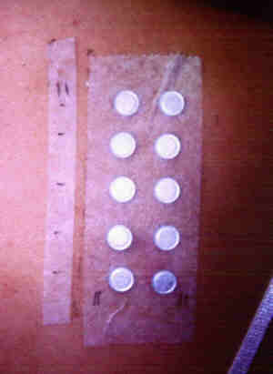
The patch test is used to identify the cause of allergic contact dermatitis. It involves applying a microporous tape with multiple aluminium chambers (called Finn chambers) containing the test chemicals to the back. After 48 hours, the tape is removed and the skin is examined 20 minutes later for a reaction. A positive reaction comprises of redness, swelling and even blistering. A second reading is carried out at 96 hours to detect delayed reactions. Patch tests are normally done only after the dermatitis is less acute, otherwise ambiguous results may be obtained.
Patch tests should be interpreted by a dermatologist because it requires skill to distinguish an allergic reaction from an irritant one. In the standard patch test, 24 or more common allergy causing chemicals are used. Additional chemicals are added depending on the history of exposure and the type of dermatitis. Once the cause has been identified, further exposure to the allergen must be avoided in order to avoid a recurrence. Patch tests only confirm or exclude an allergy to the substances tested. A negative test does not exclude an allergy because the culprit may not have been included in the test. It takes an experienced doctor and careful history taking to determine which chemicals should be added.
|
|
Patch tests. Click on image for larger view |
Photopatch tests
Photopatch tests are a special type of patch test used to detect
allergy to chemicals that occur only in the presence of sunlight.
It is done like the standard patch test above except that two
sets of chemicals are used and one set is exposed to ultraviolet
A light (UVA). It is used to diagnose photoallergic
contact dermatitis, a type of allergic contact dermatitis
that only occurs in the presence of light.
TOP
Phototests are used to detect photosensitivity (sensitivity to sunlight). It involves the use of an equipment called a monochromator which produces light of different wavelengths. One centimetre areas of skin on the back are exposed to different doses of light or different wavelengths and the test areas are examined 24 hours later for a reaction.
Photoprovocation tests involve shining the test areas daily for 3 days to provoke a reaction. They are used for the diagnosis of polymorphic light eruption (PMLE).
Photopatch tests are a special type of patch test used to detect allergy to chemicals that occur only in the presence of sunlight (photocontact dermatitis). It is done like the standard patch test above except that two sets of chemicals are used and one set that the test areas are exposed to ultraviolet A light (UVA).
These procedures are often used to treat deep ice-pick acne scars and involve the use of the biopsy punch. This instrument which works like a cookie-cutter is used to remove the core of scarred skin or a small skin abnormality. In a simple punch excision, the edges of the wound are simply stitched together. In punch grafting, a skin graft is taken from a hidden site, usually the back of the ear with the same instrument and transferred to the wound. Punch elevation is another modification that may be performed as a prelude to chemical peels, dermabrasion and laser resurfacing. In punch elevation, the core of skin is not discarded but is elevated from the underlying fat and held in place with a special tape or a stitch.
RAST is an acronym for radioallergosorbent test. It detects IgE antibodies in the blood to specific antigens (allergy causing substances). RAST can detect immediate allergic reactions to house dust, house dust mites and some food products and drugs. It gives similar information to skin tests.
In this method, a razor blade or scalpel blade is used to shave the raised abnormality flush with the skin. It is also done under local anaesthesia and does not require any suturing. It is often used for removing raised moles and seborrhoeic keratoses (age warts).
This is probably the most common procedure done to remove small abnormalities. A local anaesthetic injection is given beforehand. An elliptical incision is made around the lesion and the edges are sutured together. The incision is aligned in the direction of the normal skin lines so that the resultant scar will not be very obvious.
This is one of the most common procedures done in dermatology. It involves removing a piece of skin for histopathologic examination using a light microscope, an immunofluorescence microscope or electron microscope. The tissue may sometimes also be sent for culture (growth) of fungi, bacteria or virus.
There are four commonly used
techniques:
Skin tests, like the RAST, are used to identify the cause of immediate (IgE mediated) allergic reactions, ie., reactions that occur within minutes of exposure. Immediate allergic reactions may occur in the form of urticaria, anaphylaxis, angioedema or contact urticaria. The culprit may be a substance that is inhaled (eg., house dust, house dust mite and pollens), ingested (eg., drug or food item), injected into the body (eg., x-ray contrast media, vaccines, drugs or insect bites and stings) or that has come into contact with the skin (eg., contact urticaria). There are two types of skin tests:
The test sites are examined about 20 minutes later for a reaction. The severity of the reaction is then compared with controls (saline and histamine) to determine whether the reaction is a true allergic reaction or not.
Some individuals are so allergic such that they may develop a very severe reaction. These tests should therefore be performed in centres that have access to full resuscitation facilities.